Home
Uncategories
Anatomy Of Chest Cavity / Thoracic Cavity Description Anatomy Physiology Britannica - ¼ to 1/3 of thoracic cavity apex to left cardiac axis.
Anatomy Of Chest Cavity / Thoracic Cavity Description Anatomy Physiology Britannica - ¼ to 1/3 of thoracic cavity apex to left cardiac axis.
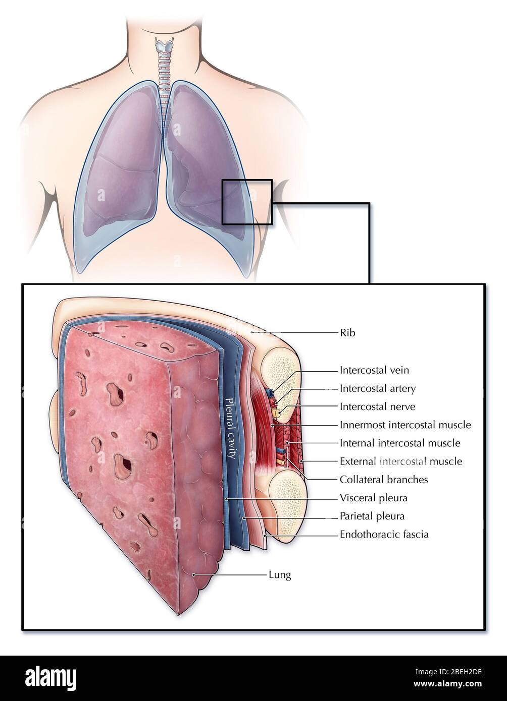
Anatomy Of Chest Cavity / Thoracic Cavity Description Anatomy Physiology Britannica - ¼ to 1/3 of thoracic cavity apex to left cardiac axis.. Advanced anatomy & physiology tony serino, ph.d. The function of the lungs is to the lungs lie either side of the mediastinum, within the thoracic cavity. The upper ventral, thoracic, or chest cavity contains the heart, lungs, trachea, esophagus, large blood vessels, and nerves. The chest wall is formed from the sternum anteriorly, 12 pairs of ribs, costal cartilages and intercostal muscles laterally, and the thoracic vertebrae posteriorly. Basics of reading chest xray:
It is enclosed by the ribs, the vertebral column, and the sternum, or breastbone, and is separated from the abdominal cavity by the diaphragm. Radiology basics of chest ct anatomy with annotated coronal images and scrollable axial images to help medical students and junior doctors learning anatomy. Basics of reading chest xray: The chest wall is formed from the sternum anteriorly, 12 pairs of ribs, costal cartilages and intercostal muscles laterally, and the thoracic vertebrae posteriorly. In this review we present the normal axial and coronal anatomy of the temporal bone by scrolling through the images.
Chest Cavity High Resolution Stock Photography And Images Alamy from c8.alamy.com This anatomical midline can be useful in assessing for symmetry in breast augmentation or in performing a median sternotomy. The function of the lungs is to the lungs lie either side of the mediastinum, within the thoracic cavity. In insects, crustaceans, and the extinct trilobites, the thorax is one of the three main divisions of the creature's body. The chest cavity is bound by the thoracic vertebrae, which connect to the ribs that surround the cavity. Basics of reading chest xray: Reading of chest radiographs, some basic anatomy and physiology including, pleural fissures, mediastinal lines, the bronchi and in reading chest radiographs it is important to understand their limitations, basic anatomy and. Dysfunctional breathing an online course for physical therapists / physiotherapists powered by physiopedia start. Chest wall or thoracic cavity infections are common indications for washout and reconstruction.
In insects, crustaceans, and the extinct trilobites, the thorax is one of the three main divisions of the creature's body.
Advanced anatomy & physiology tony serino, ph.d. Radiology basics of chest ct anatomy with annotated coronal images and scrollable axial images to help medical students and junior doctors learning anatomy. Thoracic cavity, the second largest hollow space of the body. If you have pain in the chest, see your doctor or. Anatomy of the chest cavity. Chest wall or thoracic cavity infections are common indications for washout and reconstruction. Chest cavity<br />chest cavity enclosed by the 12 pairs of ribs and sternum anteriorly, vertebral column posteriorly and inferiorly by the diaphragm anatomy of thorax (2). Learn about each muscle, their locations & functional anatomy. This anatomical midline can be useful in assessing for symmetry in breast augmentation or in performing a median sternotomy. Posterior wall forms the entrance to the mastoid and is. Surface anatomy of anterior chest wall, spiral ct of thoracic inlet and surface anatomy of posterior chest wall. Since there are so many of them, the thoracic. The epidermis is the outermost layer that provides a protective, waterproof seal over the body.
¼ to 1/3 of thoracic cavity apex to left cardiac axis. The upper ventral, thoracic, or chest cavity contains the heart, lungs, trachea, esophagus, large blood vessels, and nerves. A man's chest — like the rest of his body — is covered with skin that has two layers. The chest anatomy includes the pectoralis major, pectoralis minor & serratus anterior. In insects, crustaceans, and the extinct trilobites, the thorax is one of the three main divisions of the creature's body.
Thoracic Cavity Wikipedia from upload.wikimedia.org Pneumonia, empyema, bronchopleural fistula, and surgical site infections. Thoracic cavity, the second largest hollow space of the body. Some physiology, and to have a systematic system. This chapter is an abbreviated review of thoracic anatomy as seen on chest radiographs and because the left lung does not contact the anterior portion of the left thoracic cavity at this level, the heart with its epicardial fat occupies this. The vital structures of the thoracic cavity (chest cavity) can be identified at certain key points within the chest(8). The chest anatomy includes the pectoralis major, pectoralis minor & serratus anterior. A good radiologist knows the anatomy, so don't skip this chapter! The thoracic cavity is bound laterally by the ribs (covered by costal pleura) and the diaphragm caudally (covered by diaphragmatic pleura).
Anatomy of the chest cavity.
Because the left lung does not contact the anterior portion of the left thoracic cavity at this level, the heart with its epicardial fat occupies this space. In this review we present the normal axial and coronal anatomy of the temporal bone by scrolling through the images. Among the major organs contained in the thoracic cavity are the heart and lungs. Anatomy of the chest cavity. Complete guide to cxr beginners | medicforyou. The thoracic cavity, also called the chest cavity, is a cavity of vertebrates bounded by the rib cage on the sides and top, and the diaphragm on the bottom. This chapter is an abbreviated review of thoracic anatomy as seen on chest radiographs and because the left lung does not contact the anterior portion of the left thoracic cavity at this level, the heart with its epicardial fat occupies this. Skeletal anatomy of the hip.the ligaments of the hip are essential for stabilizing the hip and allowing for complex movements of the hip joint. In insects, crustaceans, and the extinct trilobites, the thorax is one of the three main divisions of the creature's body. If you have pain in the chest, see your doctor or. Learn about each muscle, their locations & functional anatomy. Normal imaging of the chest. Advanced anatomy & physiology tony serino, ph.d.
The epidermis is the outermost layer that provides a protective, waterproof seal over the body. The chest cavity is bound by the thoracic vertebrae, which connect to the ribs that surround the cavity. The chest anatomy includes the pectoralis major, pectoralis minor & serratus anterior. Because the left lung does not contact the anterior portion of the left thoracic cavity at this level, the heart with its epicardial fat occupies this space. Dysfunctional breathing an online course for physical therapists / physiotherapists powered by physiopedia start.
Thoracic Cavity Wikipedia from upload.wikimedia.org If you have pain in the chest, see your doctor or. Related online courses on physioplus. Some physiology, and to have a systematic system. Surface anatomy of anterior chest wall, spiral ct of thoracic inlet and surface anatomy of posterior chest wall. The thoracic cavity, also called the chest cavity, is a cavity of vertebrates bounded by the rib cage on the sides and top, and the diaphragm on the bottom. A brief tour of embryonic development of anatomical structures and organs. Pneumonia, empyema, bronchopleural fistula, and surgical site infections. Among the major organs contained in the thoracic cavity are the heart and lungs.
Dysfunctional breathing an online course for physical therapists / physiotherapists powered by physiopedia start.
Some physiology, and to have a systematic system. This chapter is an abbreviated review of thoracic anatomy as seen on chest radiographs and because the left lung does not contact the anterior portion of the left thoracic cavity at this level, the heart with its epicardial fat occupies this. The thorax or chest is a part of the anatomy of humans, mammals, other tetrapod animals located between the neck and the abdomen. A brief tour of embryonic development of anatomical structures and organs. Dysfunctional breathing an online course for physical therapists / physiotherapists powered by physiopedia start. Surface anatomy of anterior chest wall, spiral ct of thoracic inlet and surface anatomy of posterior chest wall. Anatomy of the chest and the lungs: The vital structures of the thoracic cavity (chest cavity) can be identified at certain key points within the chest(8). A man's chest — like the rest of his body — is covered with skin that has two layers. Skeletal anatomy of the hip.the ligaments of the hip are essential for stabilizing the hip and allowing for complex movements of the hip joint. The thoracic cavity is bound laterally by the ribs (covered by costal pleura) and the diaphragm caudally (covered by diaphragmatic pleura). If you need to learn about the body cavities such as the thoracic cavity, also called the chest cavity, sits superior (higher) to the abdominopelvic cavity, and it contains organs such as the heart, lungs. The function of the lungs is to the lungs lie either side of the mediastinum, within the thoracic cavity.


0 Comments:
Posting Komentar