Upper Thigh Muscles Ct Anatomy : Figure 2 from Normal MR imaging anatomy of the thigh and ... - Reviewed by mary rodts, dnp.. Muscle the lies over the frontal bone. Dummies helps everyone be more knowledgeable and confident in applying what they know. Anatomy of the muscular system. Muscles of adductor compartment of thigh and their nerve supply are as follows: This photo gallery presents the anatomy of the abdomen by means of ct (axial, coronal, and sagittal reconstructions).
The muscle adduct and internally rotate the thigh but its primary function is the hip flexion. Muscles of the posterior cervical and upper thoracic spine 1. Topographically, the muscles in this group are classed along with the lateral torso wall and upper extremity, which is due to their location as well as their genetic development based on their embryological origin. Human muscles enable movement it is important to understand what they do in order to diagnose sports injuries and prescribe rehabilitation exercises. The muscles and fasciæ of the thigh.
Hip Joint Anatomy | Bone and Spine from boneandspine.com Anatomy of the human body. Muscular compartment, bones (tibia, fibula) and muscles. The thigh is the area between the hip and the knee joint. Muscles of the posterior cervical and upper thoracic spine 1. The muscles that move the forearm are located along the humerus, which include the triceps brachii, biceps brachii, brachialis, and brachioradialis. There are few important muscles in the abdomen and pelvis. 2, vastus medialis & intermedius muscles. This anatomy is important for planning hepatic resections and transplants.
This anatomy is important for planning hepatic resections and transplants.
While the thigh muscles will be slip into the anterior, medial and posterior groups. The upper limb muscles fall into three groups. There are few important muscles in the abdomen and pelvis. The first group arise from the shoulder girdle and cross the the muscles forming the muscle mass of the posterior thigh are the hamstrings; Microscopic anatomy of skeletal muscle. Study 14 hip/upper thigh muscles flashcards from colleen k. Muscular compartment, bones (tibia, fibula) and muscles. Anatomynote.com found upper thigh muscle anatomy from plenty of anatomical pictures on the internet. The muscles and fasciæ of the thigh. Related posts of muscle anatomy of upper thigh. The muscles that move the forearm are located along the humerus, which include the triceps brachii, biceps brachii, brachialis, and brachioradialis. Muscle the lies over the frontal bone. As the cursor is moved over a particular compartment of the lower.
Muscles that move the shoulder and arm include the trapezius and serratus anterior. Anatomy of the human body. In the upper back region, the trapezius, rhomboid major, and levator scapulae muscles anchor the scapula and clavicle to the spines of several vertebrae and in addition to moving the arm and pectoral girdle, muscles of the chest and upper back work together as a group to support the vital process of. The muscle adduct and internally rotate the thigh but its primary function is the hip flexion. This is a table of skeletal muscles of the human anatomy.
Presentation1.pptx, radiological anatomy of the thigh and leg. from image.slidesharecdn.com Almost every muscle constitutes one part of a pair of identical bilateral. Anatomical structures of the lower limb (hip, thigh, knee, leg, ankle and foot) and specific regions (compartment of the lower limb) are visible on cross section of the leg : It flexes the thigh at the hip. An interactive tutorial teaching the position, actions, innervation and attachments of the rectus femoris muscle with the aid of anatomical. We think this is the most useful anatomy picture that you need. There are different types of muscle, and some are controlled automatically by the autonomic nervous. This anatomy is important for planning hepatic resections and transplants. The first group arise from the shoulder girdle and cross the the muscles forming the muscle mass of the posterior thigh are the hamstrings;
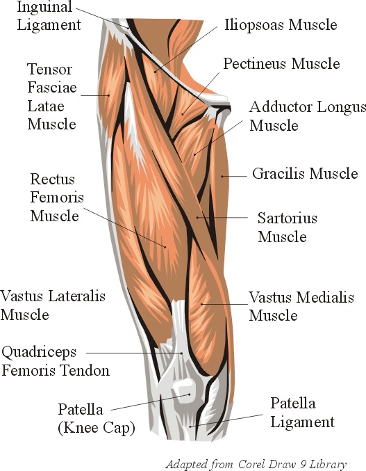
Muscles in the anterior compartment of the thigh.
It is part of the lower limb. The upper limb muscles fall into three groups. There are around 650 skeletal muscles within the typical human body. Anatomy of the muscular system. Microscopic anatomy of skeletal muscle. Muscles that move the shoulder and arm include the trapezius and serratus anterior. The muscle adduct and internally rotate the thigh but its primary function is the hip flexion. Topographically, the muscles in this group are classed along with the lateral torso wall and upper extremity, which is due to their location as well as their genetic development based on their embryological origin. Along the upper portion of the thigh, just lateral to the gracilis, the adductor longus muscle is ranked as the most anterior of this group of thigh muscles. Dummies has always stood for taking on complex concepts and making them easy to understand. Want to learn more about it? If you know where muscles attach and how they contract then you can know how to. Introduction to functional anatomy of the hip flexors and anterior thigh muscles:
Other muscles, like the skeletal muscle that moves the arm, is controlled by the somatic or voluntary nervous system. Dummies helps everyone be more knowledgeable and confident in applying what they know. Popular study materials from anatomy and cell biology 306. Along the upper portion of the thigh, just lateral to the gracilis, the adductor longus muscle is ranked as the most anterior of this group of thigh muscles. Reviewed by mary rodts, dnp.
Skeletal Muscle Review from jb004.k12.sd.us Muscular compartment, bones (tibia, fibula) and muscles. There are around 650 skeletal muscles within the typical human body. Written by keith bridwell, md; This anatomy is important for planning hepatic resections and transplants. Anatomical structures of the lower limb (hip, thigh, knee, leg, ankle and foot) and specific regions (compartment of the lower limb) are visible on cross section of the leg : For example, the quadriceps are a set of powerful muscles used to extend the leg. As the cursor is moved over a particular compartment of the lower. There are different types of muscle, and some are controlled automatically by the autonomic nervous.
Anatomy of the human body.
Regions of the upper extremity. Muscular compartment, bones (tibia, fibula) and muscles. Anatomy of the human body. An interactive tutorial teaching the position, actions, innervation and attachments of the rectus femoris muscle with the aid of anatomical. Reviewed by mary rodts, dnp. Other muscles, like the skeletal muscle that moves the arm, is controlled by the somatic or voluntary nervous system. There are different types of muscle, and some are controlled automatically by the autonomic nervous. This webpage presents the anatomical structures found on thigh mri. The upper limb muscles fall into three groups. Dummies has always stood for taking on complex concepts and making them easy to understand. Anatomy of the muscular system. Muscles are groups of cells in the body that have the ability to contract and relax. Origin is the occipital bone.
As the cursor is moved over a particular compartment of the lower upper thigh anatomy. The muscles that move the forearm are located along the humerus, which include the triceps brachii, biceps brachii, brachialis, and brachioradialis.


0 Comments:
Posting Komentar